Metabolic waste can be used to diagnose the pathophysiology of cells
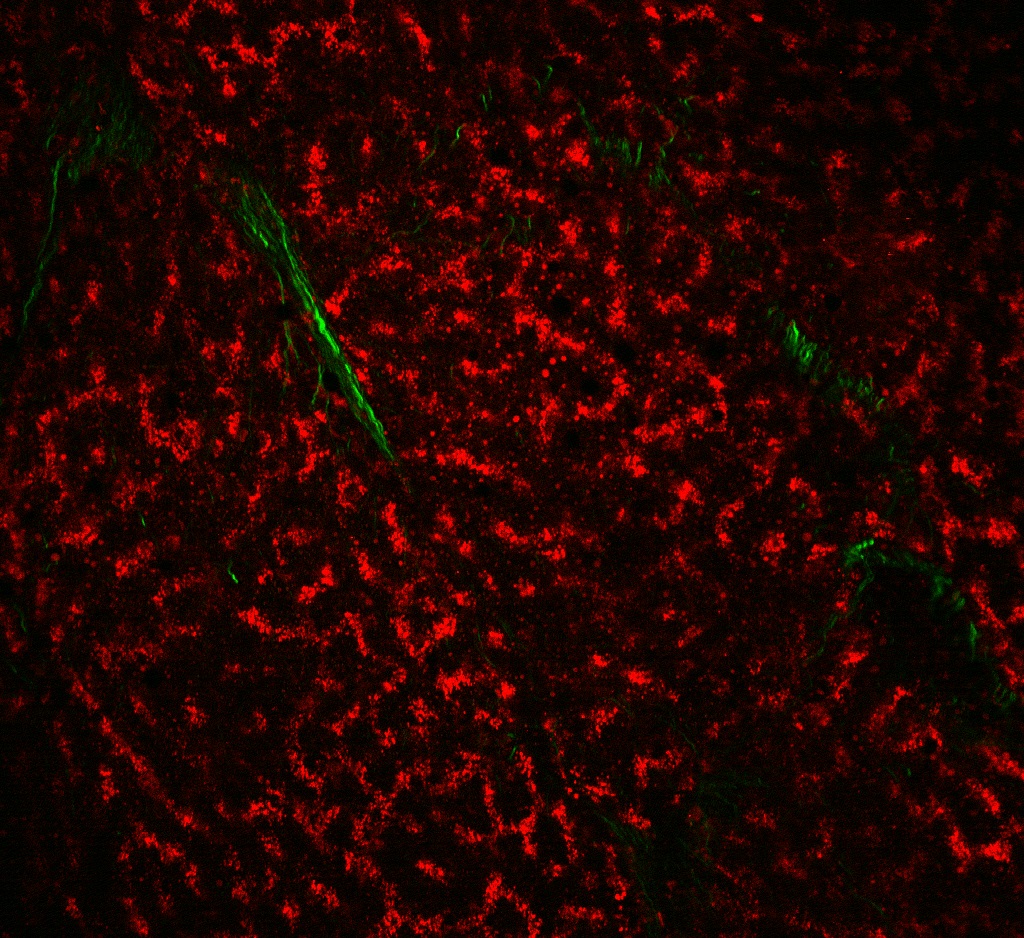
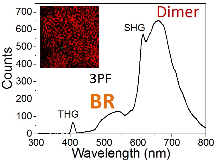
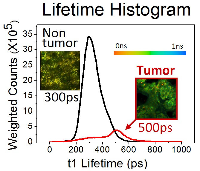
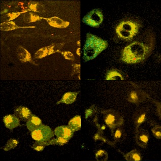
Based on an infrared femtosecond Cr:forsterite laser, we developed a semi-quantitative method to analyze the microscopic distribution of bilirubins. Using 1230 nm femtosecond pulses, for the first time, we found bilirubin dimers can emit two-photon red fluorescence around 660 nm. Autofluorescences from other endogenous fluorophores were greatly suppressed in this situation. Using this distinct fluorescence measure, we found that poorly-differentiated hepatocellular carcinoma (HCC) tissues on-average showed much lower concentration of bilirubins than the corresponding non-tumor parts. The corresponding fluorescence lifetime measurements indicated that HCC tissues exhibited a longer lifetime (500 ps) than those of non-tumor parts (300 ps). Similarly, oral cancer cell lines had longer lifetimes (>330 ps) than those of non-tumor ones (250 ps). We anticipate the developed methods of bilirubin molecular imaging to be useful in identifying the cancer boundary, diagnosing cancers, or studying the dynamics of bilirubin metabolisms in live cells.